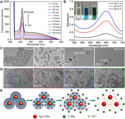
Fig. 3. Biodegradation performance.

(A) Time-dependent fluorescence spectra of Ag2S@Fe2C-DSPE-PEG dispersed in PBS buffer solution (pH 5.4, λexcitation = 370 nm, λEm = 410 nm). (B) Peroxidase-like activity of Ag2S@Fe2C-DSPE-PEG with different pH values (5.4, 6.5, and 7.4). Photo credit: Zhiyi Wang, Peking University, China. (C) TEM images (scale bars, 50 nm) of the Ag2S@Fe2C-DSPE-PEG after degradation in PBS (pH 5.4) for 0, 6, 24, and 48 hours. (D) Bio-TEM images (scale bar, 2 μm) of 4T1 cells incubated with Ag2S@Fe2C-DSPE-PEG for 24 hours (scale bars, 500 nm) of different regions enlarged. (E) Schematic representation of the degradation process of the Ag2S@Fe2C-DSPE-PEG in the physiological environment.
