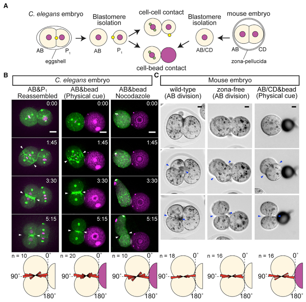
Figure 2. Physical Contact Is the Cue Sufficient for Oriented AB Cell Division in Both C. elegans and Mouse.

(A) Schematic illustration of contact reconstitution assay. Yellow dots with P1 cell indicate a midbody remnant.
(B) C. elegans AB division axis after the reconstitution of contact cues. Centrosomes (green), myosin (green), histone H2B (magenta), polystyrene bead (magenta), and cleavage furrow position (arrowheads) are shown along with the distribution of cleavage furrow orientations (bottom panel). Nocodazole 20 μg/mL was used.
(C) Mouse AB division axis in vivo and AB/CD division axis after contact reconstitution. Zona-free are embryos without zona pellucida. Cleavage furrow position (arrowheads) are shown along with the distribution of cleavage furrow orientation (bottom panel). Angle distributions are shown relative to the contact plane.
Scale bars, 10 μm. See also Figure S1.
