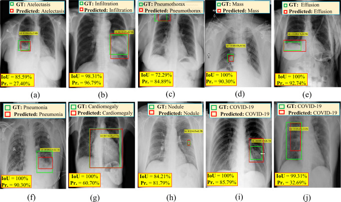
Fig. 8.
Examples of correctly predicted cases of COVID-19 and other respiratory diseases from chest X-ray (CXR) images: a atelectasis, b infiltration, c pneumothorax, d mass, e effusion, f pneumonia, g cardiomegaly, h nodule, and i & j COVID-19. The GT information (green), detected bounding box (red), IoU, and probability or confidence score (Pr.) for each case are superimposed on the original chest X-ray images

