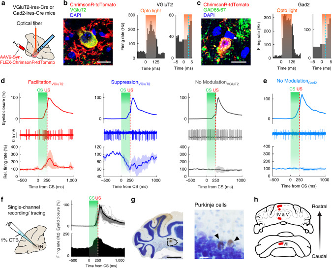
Fig. 2. Task-related modulation in excitatory FN neurons and the identification of DEC-related vermal regions.
a Schematics showing viral injection, optical fiber implantation and multichannel recording in the FN of the VGluT2-ires-Cre mice (n = 5) or the Gad2-ires-Cre mice (n = 8). b, Expression of Cre-dependent ChrimsonR in VGluT2-positive FN neurons (left), showing a short-latency response to 595 nm light (orange shading, right). The blue dashed line indicates the timing at which the firing rate exceeds three SDs of the baseline frequency within 20 ms after the light. Scale bar, 10 µm. c Same as (b), but for the Gad2-positive neurons. d Task-related modulation of VGluT2-positive neurons. Neurons are categorized based on their CS-related modulations. Top and middle rows: example eyelid movement and spike traces of individual cells. Bottom row: average firing rate of neurons with CS-related facilitation (left, n = 7), suppression (middle, n = 3) and no modulation (right, n = 5); traces are plotted as mean ± s.e.m. e Same as (d), but for the Gad2-positive neurons, showing no CS-related modulation (n = 8). f Left: experimental design for FN neuron recording and CTB tracing by using a single glass capillary. Right: a representative neuron showing CS- and US-related facilitation (overlaying eyelid closure, mean ± SD, n = 21 trials, upper right; PSTH, lower right) during the CS–US interval. g, h Iontophoresis of CTB localized to the recording site (g, left, scale bar, 1 mm) and retrogradely labeled Purkinje cells (g, right, scale bar, 20 µm) in the parasagittal vermis regions (h) (n = 5 mice).

