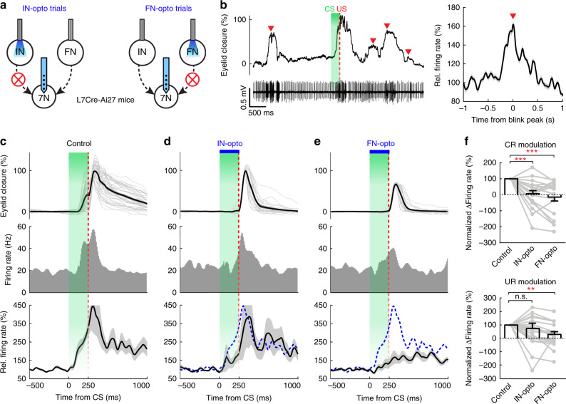
Fig. 7. Integration of FN and IN signals in 7N motor neurons synergistically controls associative behavior.
a Experimental design of 7N neuron recording during DEC from L7cre-Ai27 mice with either IN (left) or FN (right) inhibition. Arrow-headed lines indicate outputs from the IN and the FN to 7N motor neurons. b Putative 7N motor neuron activity during DEC and spontaneous eyelid movements. Left: example recording of eyelid movement (upper) and a 7N neuron (lower) showing spike rate increases in response to the DEC triggers (marked with CS and US) as well as spontaneous eyelid movements (red arrowheads). Right: average relative firing rate of all putative 7N motor neurons (n = 19, mean ± s.e.m) during spontaneous blinking (peak-aligned, red arrowhead). c–e Changes in behavior and 7N neuron activity in the control, IN-inhibition and FN-inhibition trials. Firing rate PSTH of an example 7N neuron (middle row) during behavior (upper row, n = 46, 22, 26 trials for c–e), showing that both IN and FN photoperturbations (blue bars) inhibited CR and neuron activity in response to CS, while only FN-inhibition suppressed UR and neuron activity in response to US. Lower: average spike modulation of all 7N neurons (n = 19, mean ± s.e.m). Dashed-line trace indicates the average activity during control trials in (c). f Summary of changes in relative firing rates of 7N motor neurons in response to CS and US in control, IN-opto and FN-opto trials (n = 19, mean ± s.e.m., paired two-sided t test, **P < 0.01, ***P < 0.001). CR and UR modulation (∆Firing rate) are normalized to the corresponding firing rate changes of the control session. See the exact P values for each comparison in the Source Data file.

