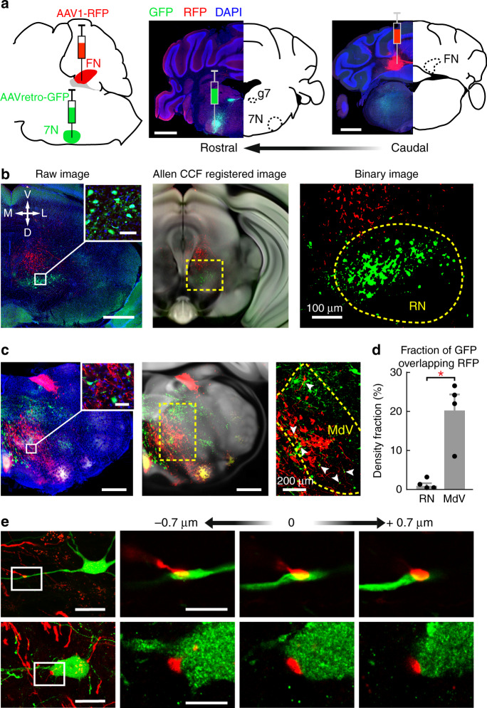
Fig. 8. Anatomical tracing reveals a FN-MdV pathway for DEC.
a Sketch of the viral tracing strategy (left). Middle and right images: coronal sections of the injection sites from an example animal. Retrograde GFP and anterograde RFP were simultaneously injected into the ipsilateral facial nucleus (7N, middle) and FN (right). Labeled fibers in the genu of facial nucleus (g7) confirm the targeting of the 7N. Scale bars, 1 mm. b Representative images of retrogradely labeled neurons (green) and anterogradely labeled FN axons (red) at the level of caudal midbrain. The raw image (left) is registered to the Allen Mouse Brain CCF (middle, see “Methods”) for further quantification of the FN projection in the contralateral red nucleus (right image, the RN is denoted by a dashed-line contour). Scale bars, 500 µm, inserted image 50 µm. c Same as (b), but for labeling in caudal medullary regions. Colocalizations of 7N projecting neurons and FN axons are found in the contralateral ventral medullary reticular nucleus (MdV, arrowheads in the right image). Scale bars, 500 µm, inserted image 50 µm. d Comparison of colocalizations in the RN and MdV from 4 mice (mean ± s.e.m., paired two-sided t test, *P = 0.020). e Confocal images of two example MdV neurons with FN axons targeting their primary dendrite (top) and soma (bottom). Scale bars, left column, 20 µm, right columns,10 µm. Tracing experiments were performed and replicated in n = 4 mice.

