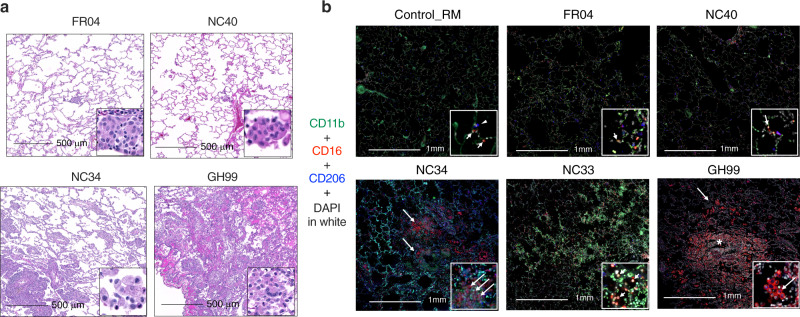
Fig. 3. Interstitial macrophages are increased in the lung of SARS-CoV-2 infected animals.
a Macrophage infiltration varied from minimal to severe in both rhesus and AGM infected with SARS-CoV-2. b Fluorescent immunohistochemistry in the lung of AGM and RM showing infiltration of CD16+ CD11b+ CD206− macrophages (arrows), with lesser numbers of CD16+ CD206+ macrophages (arrowheads). Inset showing a higher magnification of CD16+ CD11b+ CD206− macrophages (arrows). Asterix shows macrophages multifocally surrounding small airways in GH99. White: DAPI (nuclei); green: CD11b; red: CD16; blue: CD206.

