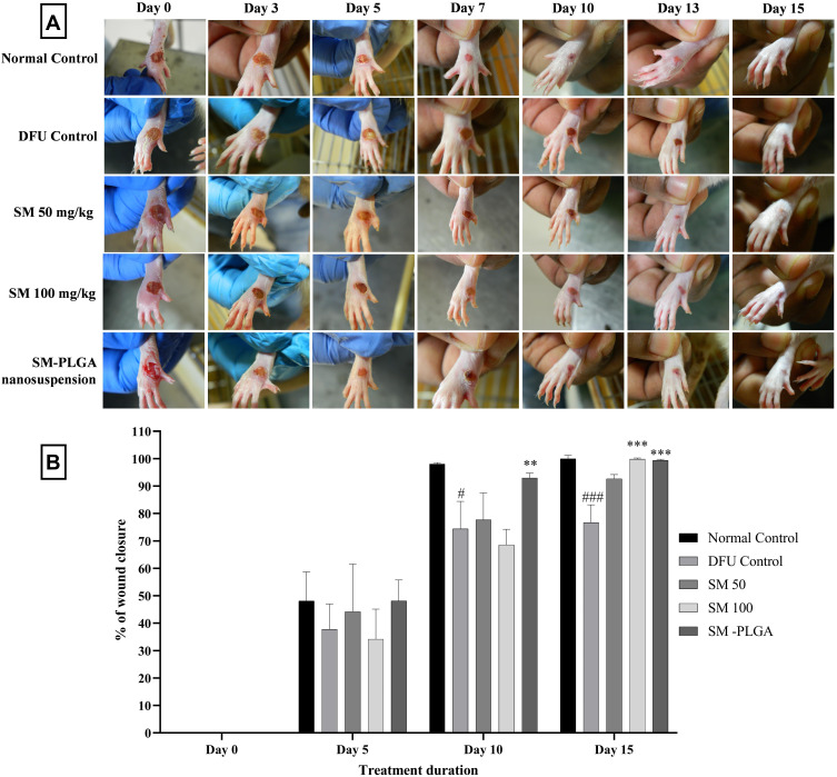
Figure 8.
(A) Representative images of DFU of treatments at 0, 3, 5, 7, 10, 13 and 15 days (B) Effect of SM-PLGA nanosuspension on wound closure in HFD+ STZ induced Type-II diabetic rats. Data represented as mean ± SEM, n=6. #p<0.05, ###p<0.001 when compared with NC, **p<0.01 and ***p<0.001 when compared with the DFU. Samples were analysed by two-way ANOVA with Bonferroni post-hoc test.

