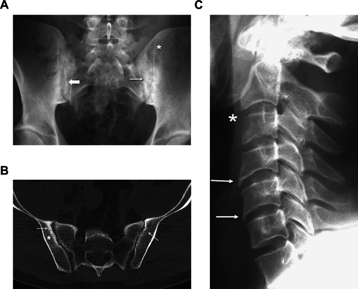
Fig. 1.
a Typical findings of sacroiliac joint involvement in a patient with ankylosing spondylitis (AS), showing extensive sclerosis (thin arrow), pseudodilation (thick arrow), and partial ankylosis (asterisk). b Computed tomography of the sacroiliac joints of a patient with AS. Note the areas with erosions (arrows) and sclerosis (asterisk), which represent characteristic signs of the disease. c Typical osteodestructive changes seen as erosion (asterisk) and osteoproliferative changes seen as syndesmophytes (arrows) in the cervical spine of a patient with AS. Reprinted from Hochberg MC, et al. Rheumatology. 7th ed.
Copyright © 2019 with permission from Elsevier

