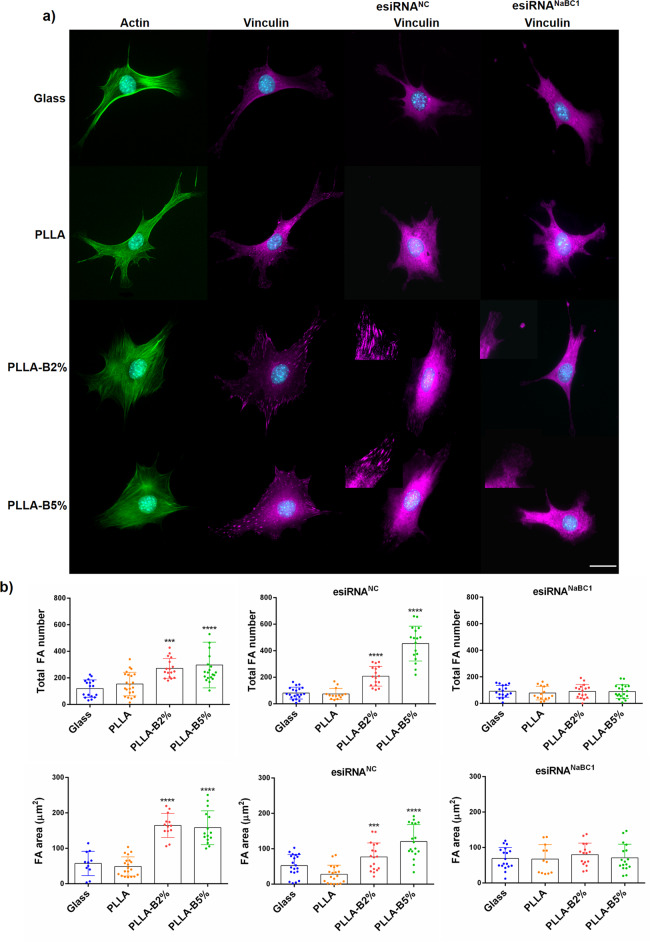
Fig. 1. Borax effects on Focal Adhesion (FA) formation and MSC adhesion.
a Immunofluorescence images of actin cytoskeleton (green), nuclei (cyan) and vinculin (magenta) as a marker of focal adhesions (FA). MSCs were cultured for 3 h, onto functionalized substrates (FN-coated), with serum-depleted and borax (PLLA-B2%, PLLA-B5%) presence in the culture medium. The same experiment was performed after NaBC1 silencing using esiRNAs (esiRNANaBC1). Universal negative control esiRNA (esiRNANC) was used as a transfection efficiency control. The PLLA-B2% and PLLA-B5% substrates presented clear focal adhesions as a result of vinculin staining that strongly diminished after NaBC1 silencing (see inset magnifications). Scale bar 25 µm. b Image analysis quantification of different parameters related to FA. Total FA number and FA area before and after NaBC1 silencing. Borax induces more and bigger FAs, effect that is reverted after NaBC1 silencing. Twenty images/condition from three different biological replicas were analyzed. The data represented in graphs correspond to n ≥ 11. Statistics are shown as mean ± standard deviation. Data were analyzed by an ordinary one-way ANOVA test and corrected for multiple comparisons using Dunnett analysis (P = 0.05). ***p < 0.001, ****p < 0.0001.

