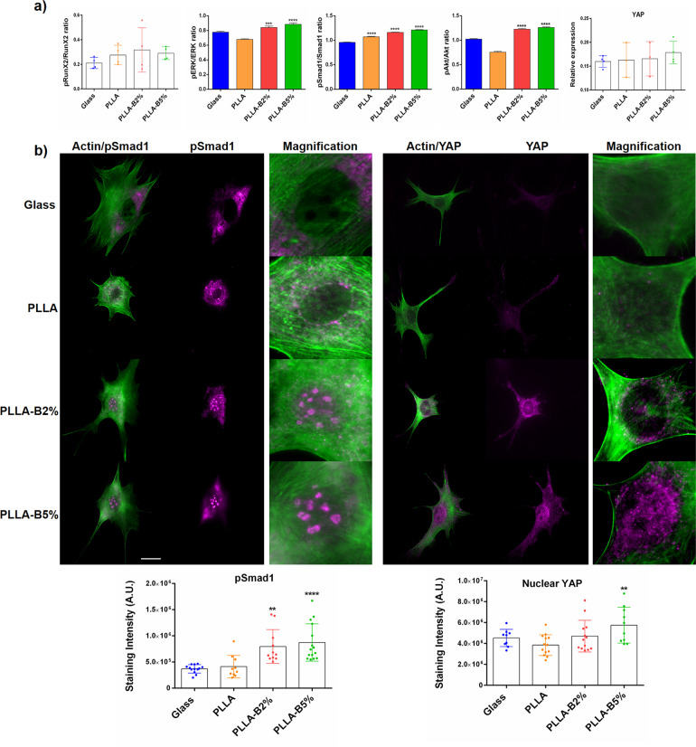
Fig. 5. Effects of NaBC1 activation in intracellular signaling.
a In-Cell Western assay showing pRunx2/Runx2, pERK/ERK, pAkt/Akt, and pSmad1/Smad1 ratios and active form of total cellular YAP on MSCs cultured onto functionalized substrates (FN-coated) serum-depleted and borax (PLLA-B2%, PLLA-B5%) in the culture medium after 90 min of culture. Simultaneous stimulation of FN-binding integrins and NaBC1 resulted in a significant enhancement of ERK, Smad1, and Akt phosphorylation, as well as active total cellular YAP detection. Graphs showing the ratio between phosphorylated/total proteins are represented as mean ± propagated standard deviation. Statistics for cellular YAP are shown as mean ± standard deviation. n ≥ 3 different biological replicas. Data were analyzed by an ordinary one-way ANOVA test and corrected for multiple comparisons using Tukey analysis (P = 0.05). *p < 0.05, **p < 0.01, ****p < 0.0001. b Immunofluorescence images of MSCs cells cultured for 3 h onto functionalized substrates (FN-coated), serum-depleted and borax (PLLA-B2%, PLLA-B5%) in culture medium. Images show actin cytoskeleton (green), pSmad1 and active YAP (magenta). Borax induces active Smad1 and YAP translocation into the cell nucleus (see inset magnifications). Scale bar 25 µm. Image analysis quantification of pSmad1 and active nuclear YAP levels. 15 images/condition from three different biological replicas were analyzed. The data represented in graphs correspond to n ≥ 9. Statistics are shown as mean ± standard deviation. Data were analyzed by an ordinary one-way ANOVA test and corrected for multiple comparisons using Tukey analysis (P = 0.05). **p < 0.01, ****p < 0.0001.

