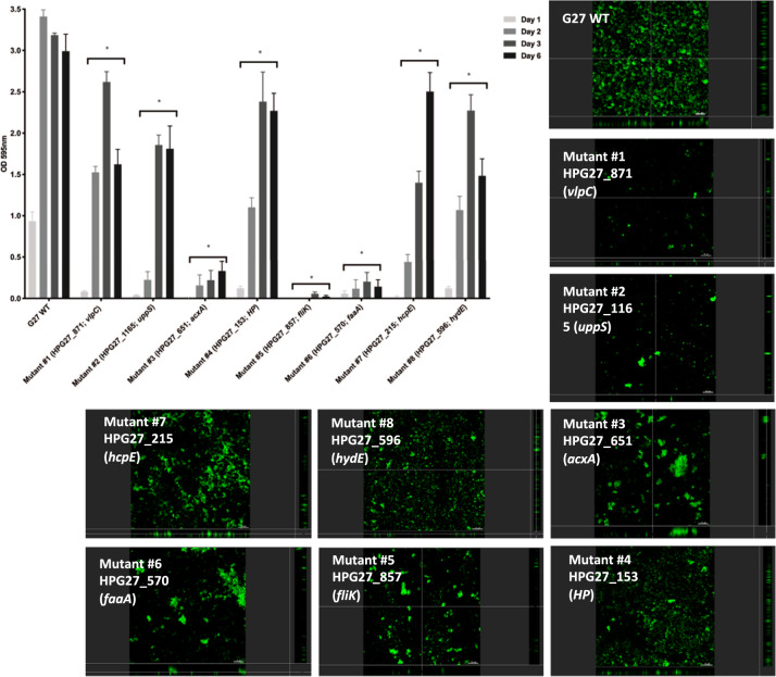
Fig. 5. Biofilm formation by H. pylori G27 WT and biofilm-defective mutants.
Biofilm formation was assessed using the microtiter plate biofilm assay and confocal laser-scanning microscopy over three days. Results represent the crystal violet absorbance at 595 nm, which reflect the biofilm biomass. Experiments were performed three independent times with at least three technical replicates for each. Error bars represent standard errors for each average value. CLSM micrographs compare biofilm formation of the wild-type G27 strain and biofilm-defective mutants. Biofilms were stained with FM 1–43, which become fluorescent once inserted in the cell membrane. HP hypothetical protein, WT Wild-type. Statistical analyses were performed using ANOVA (*P < 0.05).

