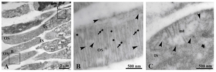
Figure 1.
Immunogold experiment with transmission electron microscopy (TEM) on bovine retina. (A) Retina section showing inner (IS) and outer (OS) photoreceptor segments. (B,C) Enlargements corresponding to the squared areas B, C in Panel A, to show a detail of an OS and mitochondrion. Largest gold particles (40 nm width, arrows) reveal Ab against anti-rhodopsin in OS. The smallest gold particle (10 nm width, arrowhead) reveals Ab against anti-ATP synthase β-subunit in both OS and a mitochondrion.

