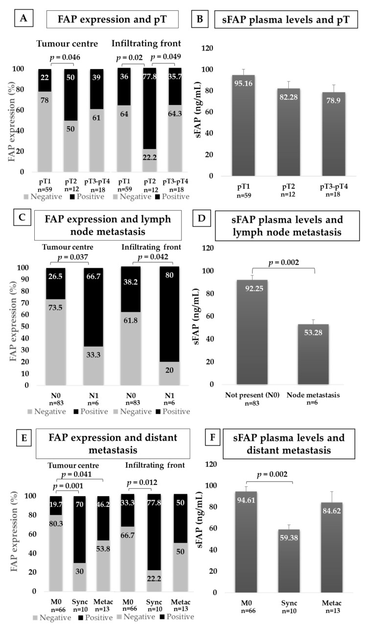
Figure 3.
Immunohistochemical FAP staining and plasma levels of sFAP according to CCRCC aggressiveness. FAP immunostaining in centre and border of tumours and sFAP levels in plasma from CCRCC patients depending on local invasion (A,B), locoregional lymph node invasion (C,D) and distant metastasis (E,F). FAP staining intensity was scored as negative or positive and Chi-square test was used for comparisons. Results in plasma samples were analysed with Mann–Whitney U-test. N0: No lymph node metastasis; N1: lymph node metastasis; M0: No distant metastasis; Sync: Synchronous distant metastasis; Metac: Metachronous distant metastasis.

