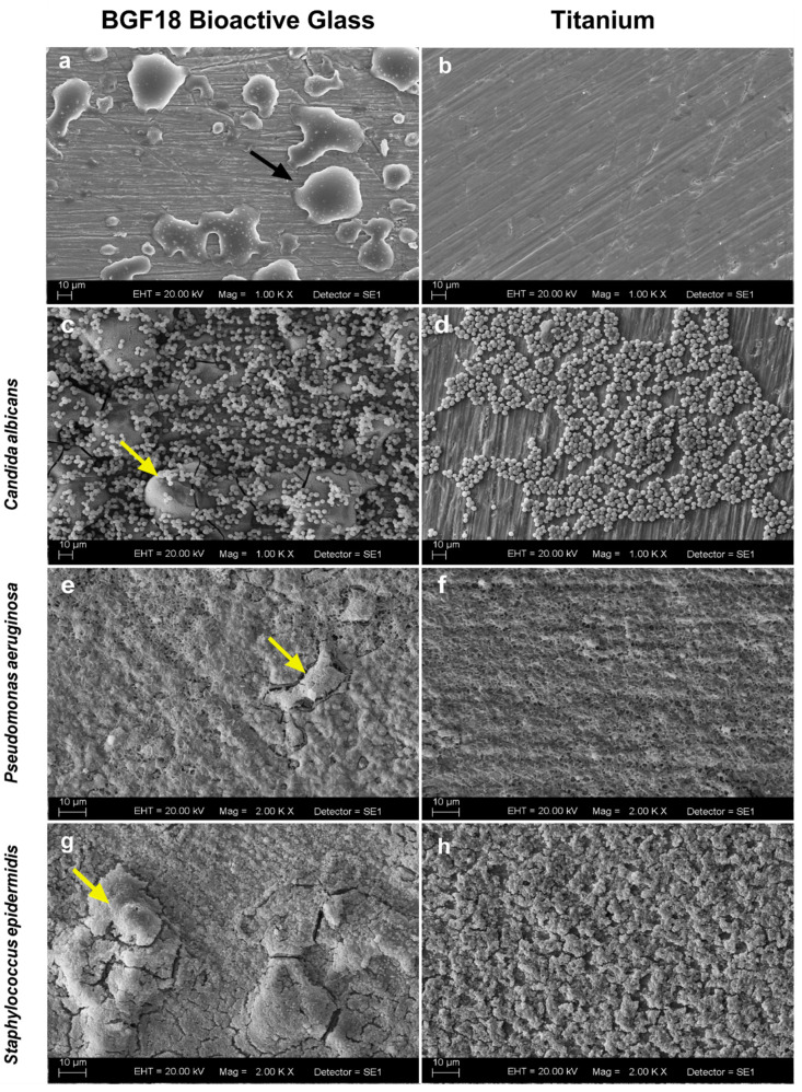
Figure 1.
(a,b) Scanning electron microscopy of a titanium specimen covered with BGF18 (magnification 1000×) and titanium (magnification 1000×), respectively. (c–h) Biofilms at 48 hours of C. albicans ((c,d), magnification 1000×), P. aeruginosa ((e,f), magnification 2000×), and S. epidermidis ((g,h), magnification 2000×), growth on titanium specimen covered with BGF18 and titanium surfaces. Black arrow indicates BGF18 deposition on the titanium surface, while yellow arrows indicate the directly colonization on the BGF18 aggregates.

