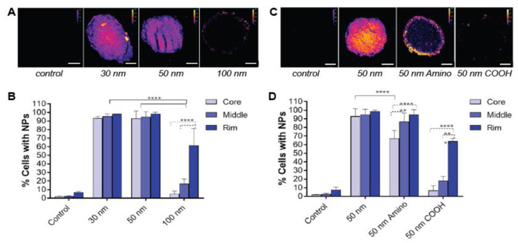
Figure 2.
The effect of nanoparticle size (A,B) and charge (C,D) on penetration depth in spheroids. (A) Confocal images of 20 µm frozen sections of HCT116 spheroids after 24 h incubation with 30, 50 and 100 nm polystyrene nanoparticles. Scale bar: 100 µm. (B) The distribution of the different sized particles (30, 50 and 100 nm) across the spheroid, distinguishing the core, middle and rim. (C) Confocal images of 20 µm frozen sections of HCT116 spheroids after incubation with 50 nm polystyrene nanoparticles (unmodified, aminated and carboxylated polystyrene nanoparticles). Scale bar: 100 µm. (D) The distribution of the 50 nm particles with different surfaces (unmodified, aminated and carboxylated) across the spheroid, distinguishing the core, middle and rim (****, ** and * indicate p < 0.0001, p < 0.01 and p < 0.05, respectively). Adapted from Reference [37], with permission of American Chemical Society © 2020.

