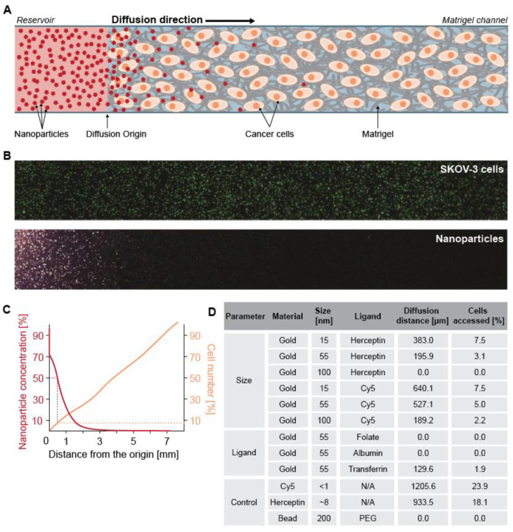
Figure 5.
(A) Scheme of nanoparticle diffusion in 3D SKOV-3 cells (ovarian cancer cell line) embedded in Matrigel® that shows impeded access of nanoparticles to the SKOV-3 cancer cells due to the Matrigel® presence. (B) Confocal images of SKOV-3 cells and nanoparticles in the Matrigel®. (C) Quantification of nanoparticle diffusion distance and the related percentage of SKOV-3 cells accessed by the nanoparticles. The diffusion distance was indicated as 50% of the initial nanoparticle concentration away from the reservoir (orange line). The corresponding distance also reflected the percentage of cells the nanoparticles had access to (orange line). (D) The average values of the diffusion distance and the percentage of accessed cancer cells the nanoparticles for other nanoparticles with various design parameters. Adapted from reference [91], with permission from American Chemical Society © 2020.

