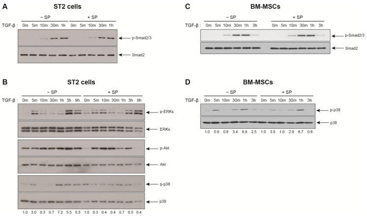
Figure 5.
The SP pretreatment decreases p38 activation in response to TGF-β. (A,B) Western blot analysis of ST2 cells that been treated 10 ng/mL TGF-β for the indicated time intervals after pretreatment with SP (+SP) or solvent (−SP). (C,D) Western blot analysis of BM-MSCs that have been treated 1 ng/mL TGF-β for the indicated time intervals after pretreatment with SP (+SP) or solvent (−SP). Protein levels of total Smad2/3, ERKs, Akt, or p38 served as the internal control for phosphorylated Smad2/3 (p-Smad2/3), phosphorylated ERKs (p-ERKs), phosphorylated Akt (p-Akt), and phosphorylated p38 (p-p38), respectively. The band intensity of p-p38 was normalized against that of p38, and the ratios are shown (two independent experiments).

