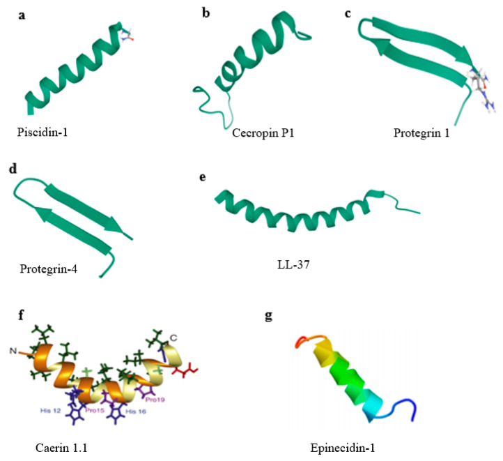
Figure 2.
Structure types of typical AMPs. (a) Solid-state NMR structure of piscidin-1 in aligned 1:1 phosphatidylethanolamine/phosphoglycerol lipid bilayers (PDB ID 2MCV). (b) Solution structure of cecropin P1 with LPS (PDB ID 2N92). (c) Protegrin 1 from porcine leukocytes, NMR, 20 structures (PDB ID 1PG1). (d) Structure of protegrin-4 by high-resolution NMR spectroscopy (PDB ID 6QKF). (e) Structure of human LL-37 (PDB ID 2K6O). (f) Solution structures of caerin 1.1 [56]. (g) Structure of epinecidin-1 [57].

