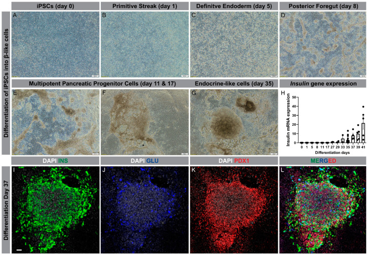
Figure 2.
Differentiation of human iPSCs into β-like cells. (A–G): Progression through different developmental stages during differentiation of iPSCs into β-like cells (scale bar is 200 µm); (H): Insulin gene expression through the course of differentiation, ranging from day 0 to day 41; (I–L): Condensed cluster of differentiated cells at day 37 displaying protein expression of insulin (green), glucagon (blue), and PDX1 (red); nucleus (grey) (Scale bar is 50 µm).

