Figure 2.
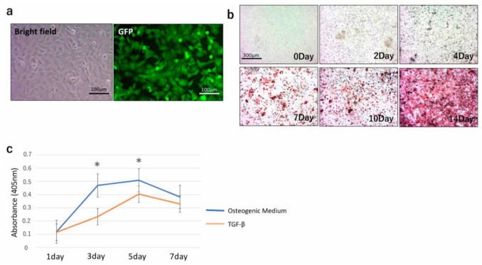
(a) TGC showed a fibroblastic shape (left), and fluorescent microscopy confirmed the expression of green fluorescent protein (GFP) (right). (b) Alizarin red staining of TGC exposed to osteogenic medium from 0 to 14 days. (c) Measurement of Alkaline Phosphatase (ALP) activity of TGC cultivated with osteogenic medium or TGF-β. The ALP activity from osteogenic medium became significantly higher than that from TGF-β at 3 and 5 days. After this point, the ALP activity of osteogenic medium- and TGF-β-exposed TGC decreased. * p < 0.05.

