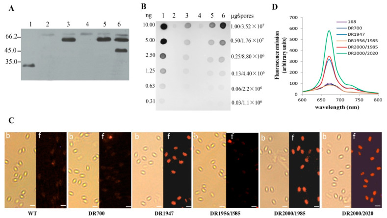
Figure 2.
Detection of the Vp7 antigen in the recombinant B. subtilis spores. (A) Western blot analysis of the Vp7 antigen in the spore coat proteins. Lane 1, the purified His6-Vp28; lanes 2–6, the coat proteins extracted from the spores of the recombinant strains DR700, DR1947, DR1956/1985, DR2000/1985, and DR2000/2020, respectively. Molecular weight markers (kDa) are indicated at the right. (B) Dot blot analysis for the amount of the Vp7 antigen in the recombinant spore coat. The amounts of His6-Vp7 (lane 1) and the coat proteins from the spores of DR700 (lane 2), DR1947 (lane 3), DR1956/1985 (lane 4), DR2000/1985 (lane 5), and DR2000/2020 (lane 6) were indicated. (C) Immunofluorescent detection of the Vp7 antigen on the spore surface. The immunofluorescence-labeled spores were visualized under bright field (b) and fluorescence (f) microscopy. Scale bars indicate 1 μm. (D) Quantitative analysis of the Cy5 fluorescence on the spore surface with fluorospectrophotometer.

