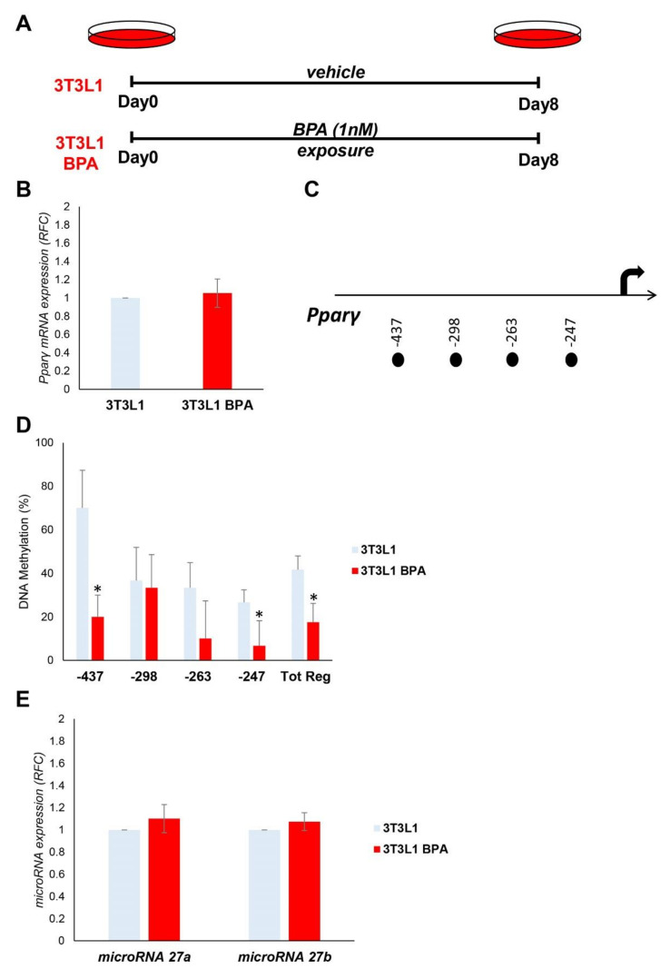
Figure 1.
BPA exposure effects on the expression and epigenetic modifications of the Pparγ gene in 3T3L1 preadipocytes. (A) Graphical representation of the 3T3L1 cells chronically exposed (8 days) to low BPA concentrations (1 nM). (B) Pparγ mRNA levels were assessed in 3T3L1 cells exposed to BPA by qPCR. Results are the mean ± SD (n = 6). (C) Schematic representation of the four CpG sites located within the 500 bp upstream of the transcription start site (TSS) of the Pparγ gene, where the methylation was investigated. (D) Bisulfite sequencing analysis for DNA methylation assessment of the Pparγ promoter region in 3T3L1 cells exposed or not to BPA. Results are the mean ± SD (n = 3); for each experiment, at least ten clones were analyzed by Sanger sequencing. (E) BPA effects on the microRNA 27a and microRNA 27b expression in 3T3L1 cells. MicroRNA 27a and microRNA 27b expression was analyzed by qPCR and quantified as expression units relative to U6 snRNA used as housekeeping small RNA. Results are the mean ± SD (n = 5). Statistical significance was tested by the two-tailed Student’s t-test. Significant p values are indicated as * p < 0.05.

