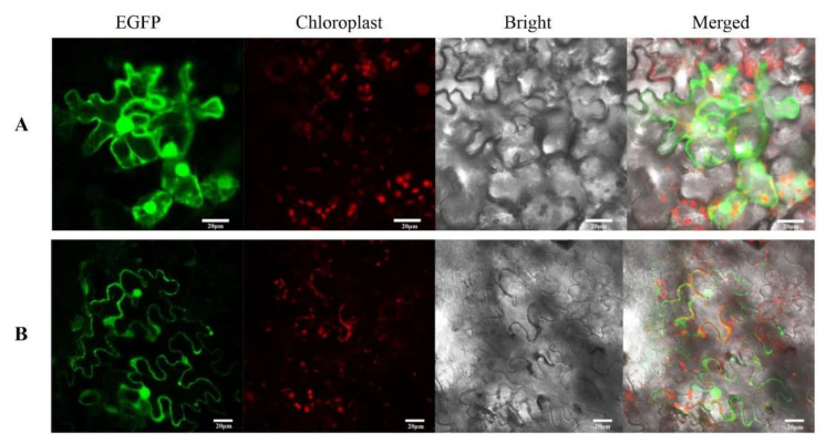
Figure 4.
The subcellular localization of CnDFR. (A) Observation by LSM510 Meta of the EGFP empty vector. White scale: 20 µm. The green fluorescence signals appeared in the nucleus, cell membrane and cytoplasm under the excitation of the wavelength of 488 nm. (B) Observation of the lower epidermal cells of Nicotiana benthamiana leaves with the CnDFR-EGFP vector. White scale: 20 µm. The nucleus and cell membrane expressed a strong green fluorescence signal.

