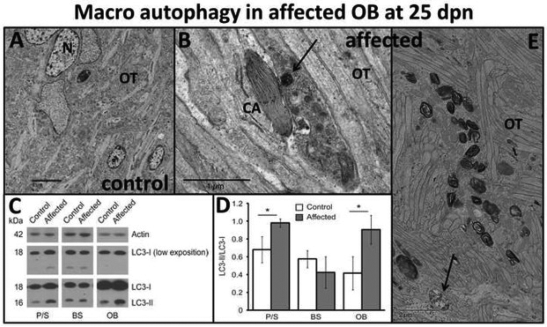
Fig. 10.

Olfactory tract (OT) pathological alterations. (A) Normal ultrastructural sagittal section through olfactory tract (OT) projections and neurons (N) in control rats at 25 dpn and (B, E) ultrastructural sagittal section showing axonal accumulation of macro autophagosomes (B, black arrow) in different degrees of development in the OT of affected rats at 25 dpn. (B) Also, adjacent there is an axon containing smooth endoplasmic reticulum in a crystalline-like configuration (CA). Left lower corner in E represents a typical early autophagosome consisting of accumulation of organelles and lysosomes surrounded by a capsule (arrow). [Bar = 5 μm (A), Bar = 1 μm (B), Bar = 2 μm (E)]. Autophagosome biomarkers: (C) LC3I to LC3II conversion. Representative immunoblots of protein extracted from putamen/striatum (P/S), brain stem (BS) and olfactory bulb (OB) of control and affected rats (25 dpn), probed with antibodies (lower exposure of LC3-I were used for quantification). (D) Graph shows the densitometric analysis expressed as the LC3-II/LC3-I ratio. Data is shown as mean ± S.E.M. *p < .05 (n = 3 animals for each experimental condition).
