Fig. 2.

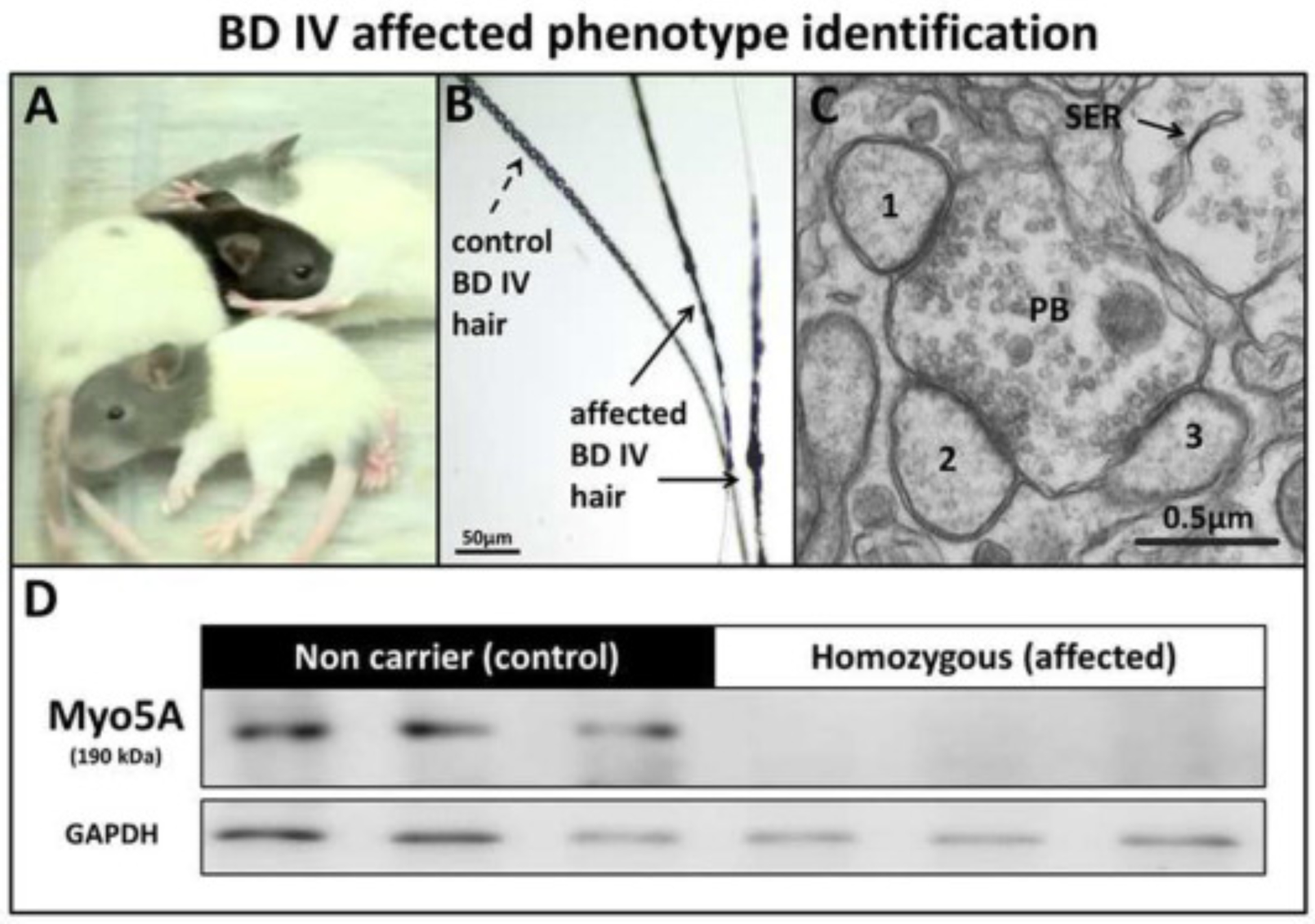
BD-IV affected phenotype identification. (A) Gray color of the head of affected rats compared with the control (non-affected littermate), (B) Accumulation of melanosomes within the hair shaft (arrows), (C) Ultrastructural cross-sectional profile of a single cerebellum large parallel fiber bouton (PB) making contact with 1, 2, 3 Purkinje cell (PC) spines from the molecular layer, showing absence of smooth endoplasmic reticulum (SER) in affected rats at 25 dpn (Bar = 0.5 μm). (D) Western blot showing loss of Myo5a protein in the brain of affected rats compared to control rats at 30 dpn.
