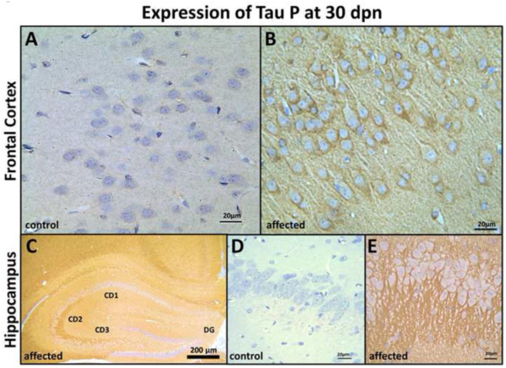
Fig. 7.

Frontal cortex (FC) and Hippocampus (HC) tau-P expression in control and affected rats at 30 dpn. FC immunohistochemistry in control rats (A) showing weak tau-P immunolabeling, and affected rats (B) showing strong labeling of axonal, perikaryal localization of tau protein (Bar = 20 μm). (C) Hippocampal formation showing strong immunolabeling of CD2 and CD3 region in the affected rats, but there is no labeling of dentate gyrus (DG) and less labeling of the CD1 region (Bar = 200 μm). Immunohistochemistry in control rats (D) showing weak tau-P immunolabeling and (E) pyramidal neurons axonal and perikaryal showing strong labeling for tau-P protein in the affected rats. (Bar = 20 μm).
