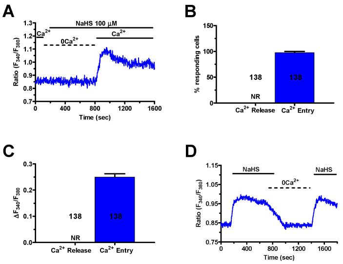
Figure 4.
Extracellular Ca2+ entry mediates the Ca2+ response to NaHS. (A), NaHS (100 μM) failed to elicit intracellular Ca2+ levels when the cells were bathed in the absence of extracellular Ca2+ (0Ca2+), the [Ca2+]i raised following re-addition of extracellular Ca2+. (B), mean ± SE of the percentage of mCRC cells presenting a discernible Ca2+ release or Ca2+ entry in response to 100 μM NaHS. (C), mean ± SE of the amplitude of Ca2+ release and Ca2+ entry induced by NaHS in mCRC cells. (D), removal of extracellular Ca2+ (0Ca2+) during the plateau phase caused the [Ca2+]i to undergo a reversible decline to pre-stimulation levels.

