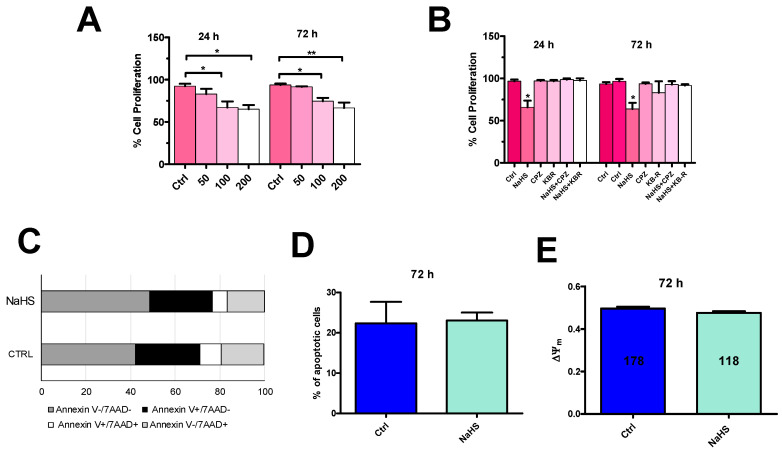
Figure 13.
NaHS inhibits proliferation, but does not induce apoptosis, in mCRC cells. (A), incubation of mCRC cells for 24 h and for 72 h in presence of increasing concentrations of NaHS (50, 100 and 200 µM) reduced cell proliferation as determined by direct cell counting with the Trypan blue assay. (B), the anti-proliferative effect of NaHS (100 µM, 72 h) was prevented by pretreating mCRC cells with capsazepine (CPZ; 10 µM, 20 min) and KB-R 7943 (KB-R; 10 µM, 30 min). One-way ANOVA analysis followed by the post-hoc Dunnett’s test was used for Statistical comparison. In Panels A and B: ** p ≤ 0.001; * p ≤ 0.05. Each experiment was repeated three times. (C), Flow cytometry analysis of Annexin and 7-ADD staining. Representative cytogram of three independent experiments with the percentage of distribution of different population of mCRC cells in the absence (Ctrl) and in the presence of NaHS (100 µM, 72 h). Living cells (Annexin V−/7−ADD−) are represented in dark grey, early apoptotic cells (Annexin V+/7−ADD−) in black, late apoptotic/dead cells (Annexin V+/7−ADD+) in white, and dead cells (Annexin V+/7−ADD+) in light grey. (D), mean ± SE of the percentage of apoptotic mCRC cells in the absence (Ctrl) and presence of NaHS. (E), mean ± SE of ΔΨm in the absence (Ctrl) and presence of NaHS (100 µM, 72 h). ΔΨm was measured by evaluating tetramethylrhodamine, methyl ester (TMRM) fluorescence [67].

