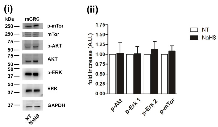
Figure 14.
NaHS does not activate phosphorylation cascades in mCRC cells. Phosphorylation of the indicated selected signaling proteins in mCRC cells incubated with 100 µM NaHS for 24 h. Representative immunoblots with specific anti-phosphoprotein antibodies directed against the different substrates are reported (i). Akt, Erk and mTOR staining were used for equal loading control for corresponding specific phosphoprotein. GAPDH staining was used as equal loading control. Quantification of the results performed by densitometric scanning is reported in (ii), as fold increase (A.U.) of phosphorylation over basal (NT). Results are the mean ± SD of three different experiments.

