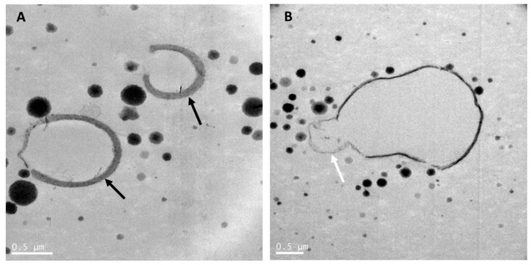
Figure 1.
Transmission electron microscopy images of raw goat milk. (A) showing goat milk caseins and fat globules with a thick MFGM. (B) showing a milk fat globule with a cytoplasmic crescent (white arrow). Black arrows point at the MFGM surrounding fat globules. Goat milk was collected from a single goat in a New Zealand goat farm and kept at room temperature until processing within 2 h using the same method as in Gallier et al. [5]. Scale bar = 0.5 µm.

