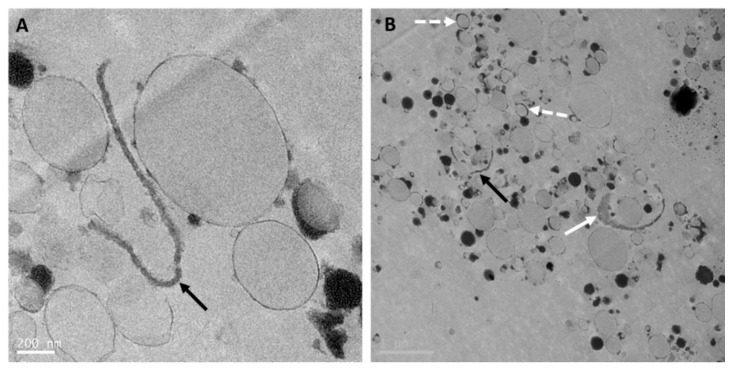
Figure 3.
Transmission electron microscopy images (A and B) of reconstituted whole goat milk-based infant formula (Dairy Goat Co-operative, Hamilton, New Zealand) processed using the same method as in Gallier et al. [5]. Black arrows point at MFGM fragments in the serum phase (A and B). Full white arrow points at an MFGM fragment with attached cytoplasmic crescent filled with electron-dense material (B). Dashed white arrows point at droplets with thicker interface (B). Scale bars = 200 nm (A) and 1 µm (B).

