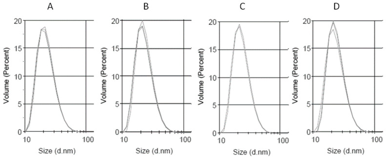
Figure 3.
Dynamic light scattering (DLS) measurement of the size distribution of the obtained low-density lipoprotein (LDL) preparations. (A) LDL, (B) QLDL, (C) CurLDL, and (D) CLALDL. PPs-doped LDL samples were prepared by mixing 0.5 mg of PPs per 1 mL of native LDL preparation (6 mg/mL of protein), followed by 11 h incubation at RT, with shaking in the dark. Three repetitions of measurements are overlaid for each preparation. The full diagrams in the range 0.1–10,000 nm have been included in Supplementary Material, Supplementary Figures S1–S4.

