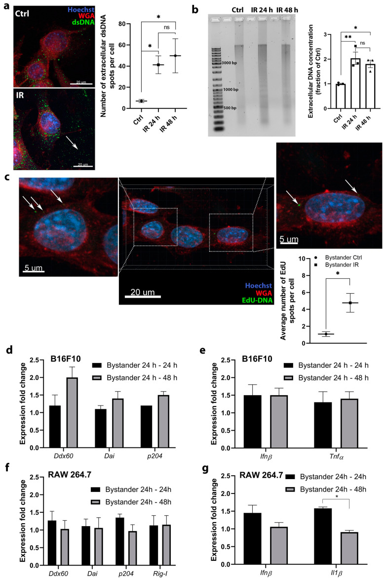
Figure 5.
Radiation induced the release of DNA into the cell medium, which entered fresh nonirradiated cells but did not induce the upregulation of DNA sensing pathways and cytokines. (a) DNA staining demonstrated that DNA is present outside the irradiated B16F10 cells in a larger amount than control cells; scale bar = 20 μm; n = 8–9 (number of quantified images). DNA was detected using anti-dsDNA antibodies; the signal is presented by arrows indicating green spots. Ctrl—control cells, IR—6 Gy irradiated cells. (b) Agarose gel analysis and measured concentration of isolated DNA from the cell culture medium from control nonirradiated cells (Ctrl) and after 6 Gy irradiation at 24 h (IR 24 h group) and 48 h (IR 48 h group); n = 3. The original image of AGE gel shown in Figure S4. (c) The detection of the uptake of DNA with incorporated EdU (arrow) from the cell-conditioned medium of irradiated B16F10 cells into the fresh nonirradiated B16F10 cells. The positive signal on images is presented by arrows indicating green spots and the quantification of images on graph representing average number of EdU spots per cell; scale bar = 20 μm and 5 μm (inset); n = 5 (number of quantified images). (d) Expression fold change of specific DNA sensors after addition of cell-conditioned medium from 6 Gy irradiated B16F10 cells to fresh nonirradiated B16F10 cells and 24 h incubation (Bystander 24 h–24 h) or 48 h incubation (Bystander 24 h–48 h); n = 4–7. (e) Expression fold change of specific cytokines after addition of cell-conditioned medium from 6 Gy irradiated B16F10 cells to fresh nonirradiated B16F10 cells and 24 h incubation (Bystander 24 h–24 h) or 48 h incubation (Bystander 24 h–48 h); n = 3. (f) Expression fold change of specific DNA sensors after addition of cell-conditioned medium from 6 Gy irradiated B16F10 cells to fresh nonirradiated RAW 264.7 macrophages and 24 h incubation (Bystander 24 h–24 h) or 48 h incubation (Bystander 24 h–48 h), n = 3. (g) Expression fold change of specific cytokines after addition of cell-conditioned medium from 6 Gy irradiated B16F10 cells to fresh nonirradiated RAW 264.7 macrophages and 24 h incubation (Bystander 24 h–24 h) or 48 h incubation (Bystander 24 h–48 h); n = 3. For panels (d–g), the values were normalized to the pertinent control groups i.e., cells to which the cell-conditioned medium of control nonirradiated cells was added. Statistical significance was determined by one-way ANOVA followed by a Dunnett’s multiple comparisons test. * p < 0.05, ** p < 0.01 vs. Ctrl. n = number of biological replicates in (b,d–g).

