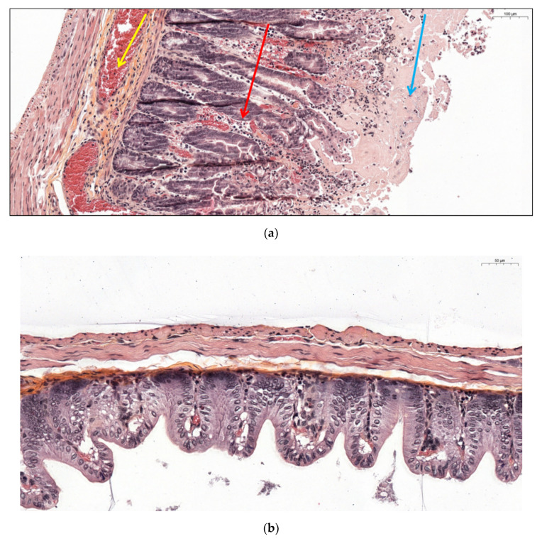
Figure 7.
(a) Caecal histological section from a hamster co-infected by PCD130 + 027 (group D) euthanized after presenting CDI clinical signs. Diffuse hemorrhage is designated by the yellow arrow, polynuclear infiltration by the red arrow, hyperplasia and epithelial desquamation by the blue arrow. (b) Intestinal histological section from a hamster colonized by PCD130 (group B) surviving at D20.

