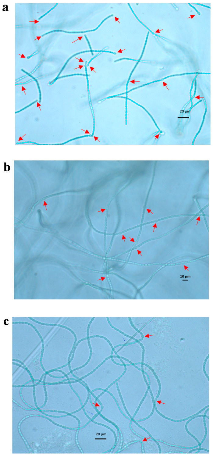
Figure 2.
Microscopic photos of A. variabilis trichomes under 400× magnification. Heterocysts shown in the photos were formed during the experiment with N-free medium (a), low nitrate (1.5 × 10⁻⁵ g L−1 of NaNO3) (b) and ammonium (0.006 × 10⁻² g L−1 of Fe-NH4-citrate) supplements (c) on day 4 when the highest heterocyst frequencies were measured. Heterocysts were able to be distinguished with adjacent vegetative cells by roundish shape and pale color. Arrows in figures indicate heterocysts.

