Figure 6.
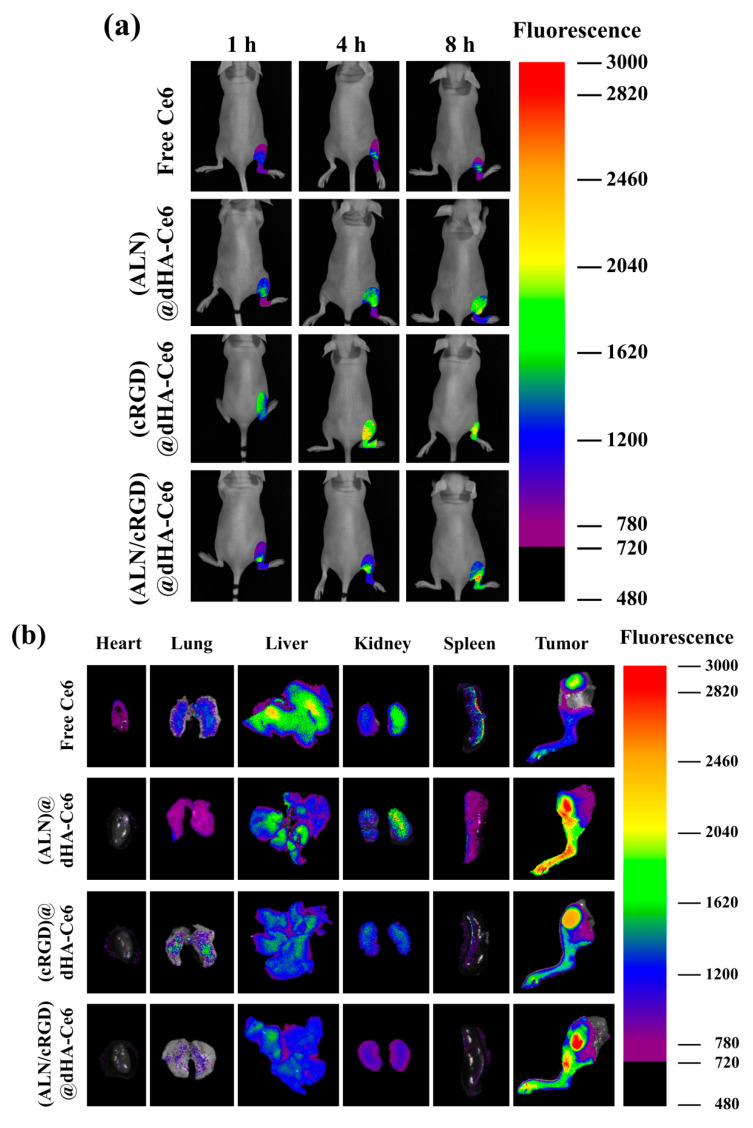
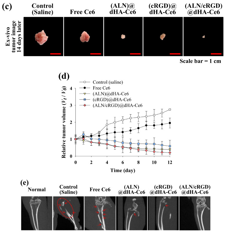
(a) In vivo noninvasive photoluminescent tumor imaging of free Ce6 (2.5 mg/kg) or each sample (equivalent Ce6 2.5 mg/kg) intravenously injected into MDA-MB-231 tumor-bearing nude mice. Fluorescent tumor images were obtained at 1, 4, and 8 h post-injection. (b) Ex vivo fluorescence images of major organs and tumors at 24 h post-injection. (c) Optical images of ex vivo tumors extracted from MDA-MB-231 tumor-bearing nude mice at 14 days post-injection of saline, free Ce6 (2.5 mg/kg), or each sample (equivalent Ce6 2.5 mg/kg). (d) Relative tumor volume change (Vt/V0, where Vt is the tumor volume at a given time and V0 is the initial tumor volume) of MDA-MB-231 tumor-bearing nude mice treated with saline, free Ce6 (2.5 mg/kg) or each sample (equivalent Ce6 2.5 mg/kg). (e) In vivo X-ray CT images of MDA-MB-231 tumor-bearing nude mice treated with control (saline), free Ce6 (2.5 mg/kg), or each sample (equivalent Ce6 2.5 mg/kg). The tumor site is indicated by a dashed circle and arrow.


