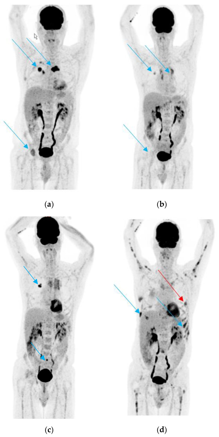
Figure 1.
(a). 18F-fluorodeoxyglucose positron emission/computed tomography (18FDG-PET/CT) scan performed before start of treatment; the arrows show the primary tumor in the lung and some of the bone metastases. (b). 18FDG-PET/CT scan performed after 3 months of treatment with erlotinib and bevacizumab. The arrows show partial remission of the primary tumor and the bone metastases. (c). 18FDG-PET/CT scan performed after 9 months of treatment with erlotinib and bevacizumab. The blue arrows show the slight progression of the primary tumor and in the sacrum. (d). 18FDG-PET/CT in January 2019. The blue arrows show new diffuse bone metastasis of several skeletal regions and the red arrow a new pleural lesion, which was biopsied.

