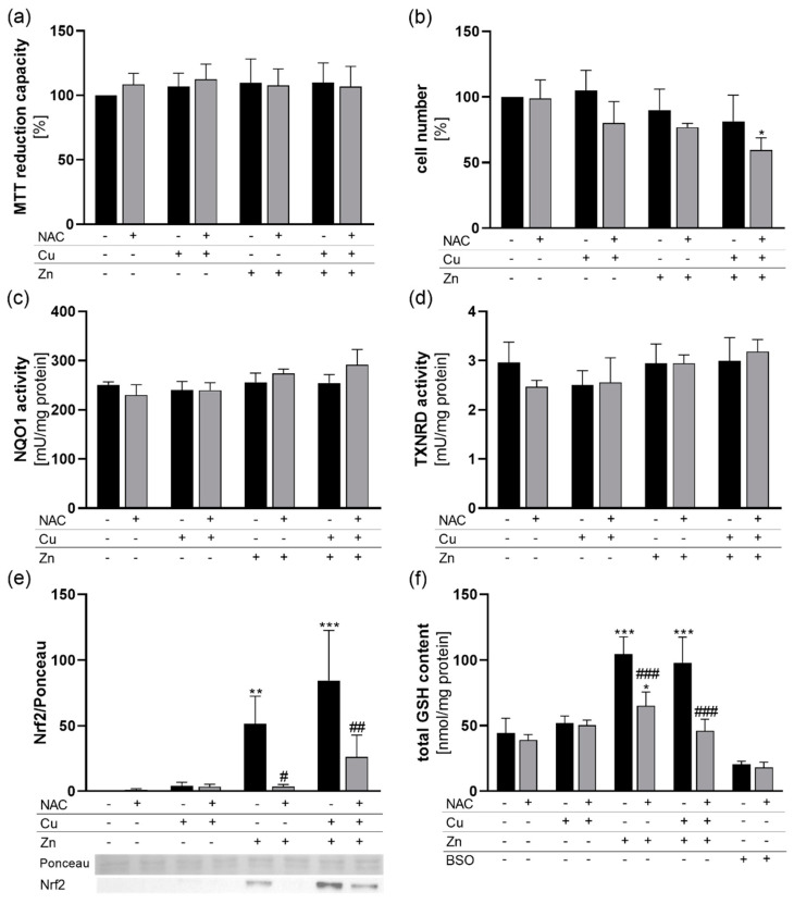
Figure 1.
Cell viability and cellular redox homeostasis in response to NAC treatment in combination with Cu and Zn. HepG2 cells were cultured for 6 (a–d,f) or 4 h (e) with or without 1 mM NAC in combination with 50 µM CuSO4, 50 µM ZnSO4 or both. (a) MTT reduction capacity and (b) cell numbers were analyzed. (c) NQO1 and (d) TXNRD activities were measured photometrically. (e) Nuclear translocation of Nrf2 was analyzed by Western blot and normalized to Ponceau staining. (f) The total GSH content of the cells was determined photometrically. The GSH synthesis inhibitor BSO was used as positive control. Results are depicted as mean + SD (n = 4). Statistical analysis was calculated by two-way ANOVA with Bonferroni´s post-test. * p < 0.05; ** p < 0.01; *** p < 0.001 vs. cells without Zn and Cu treatment (trace element effect); # p < 0.05; ## p < 0.01; ### p < 0.001 vs. −NAC (NAC effect).

