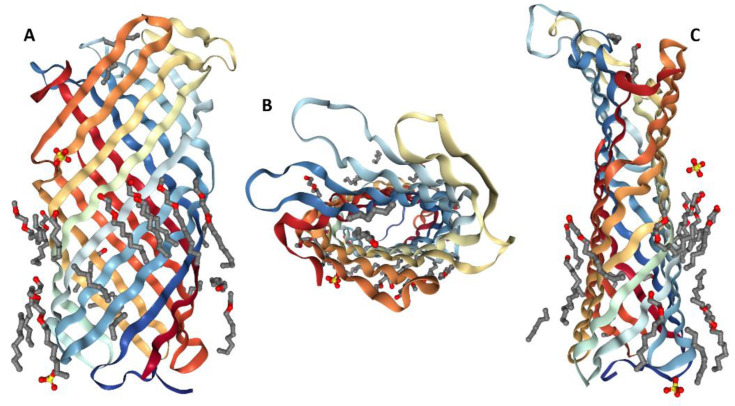
Figure 1.
3D structure of the plasminogen activator Pla from Yersinia pestis determined by X-ray crystallography (PDB ID 2 × 55) [30]. Two side projections (plots A,C) and a top view from the extracellular side (plot B) of Pla are shown together with the set of C8E4 detergent molecules.

