Figure 2.
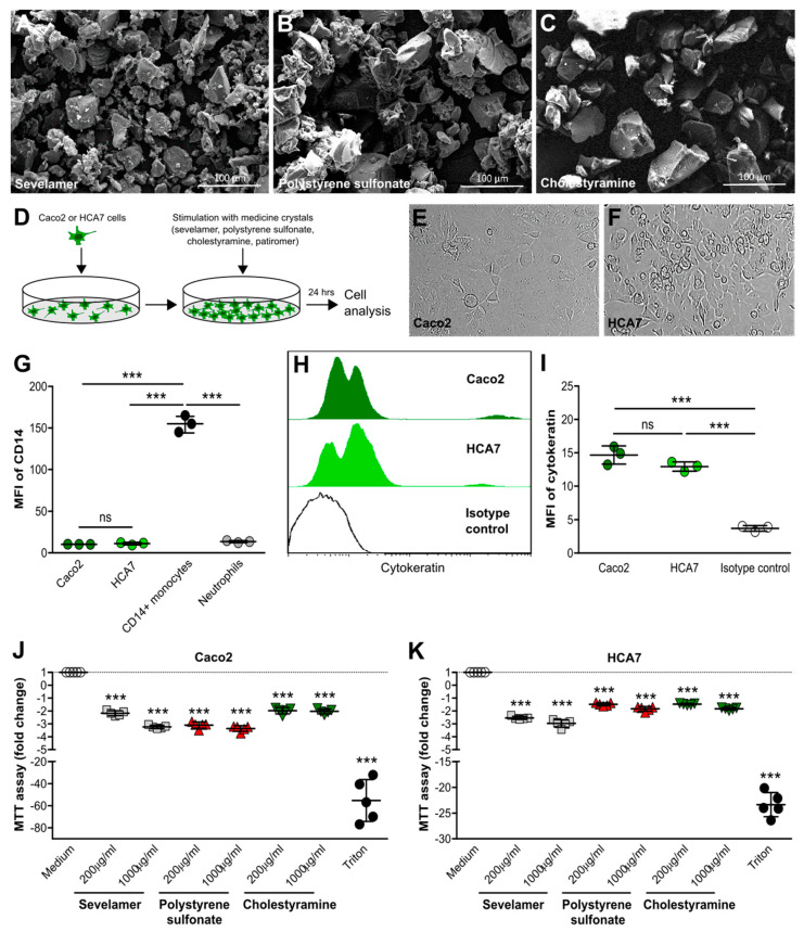
Crystals of ion-exchange resins reduce metabolic activity in intestinal epithelial cells. (A–C) Scanning electron microscopy illustrates sevelamer (A), polystyrene sulfonate (B), and cholestyramine (C) crystals of different sizes and shapes. (D) Schematic of in vitro culture set-up of human Caco2 and HCA7 intestinal epithelial cell lines. (E,F) Light microscopy of human Caco2 (E) and HCA7 (F) intestinal epithelial cell lines (400× magnification). (G) Caco2 and HCA7 cells, as well as human blood neutrophils and CD14+ monocytes, were stained for the surface marker CD14 and analyzed by flow cytometry (n = 3). (H,I) Caco2 and HCA7 cells with cytokeratin or isotype control (using CaCo2 cells) and flow cytometry performed. Histogram (H) and the mean fluorescence intensity (MFI) (I) are shown (n = 3). (J,K) Human Caco2 and HCA7 intestinal epithelial cell lines were stimulated with different concentrations of sevelamer, polystyrene sulfonate, and cholestyramine crystals (200 or 1000 µg/mL) for 24 h. Triton served as a positive control. After stimulation, culture supernatants were collected and 3-(4,5-dimethylthiazol-2-yl)-2,5-diphenyltetrazolium bromide (MTT) assays were performed. The fold change is shown (n = 5). Data are means ± SD. * p ≤ 0.05; ** p ≤ 0.01; *** p ≤ 0.001; ns, not significant (p > 0.05) versus the medium using one-way ANOVA.

