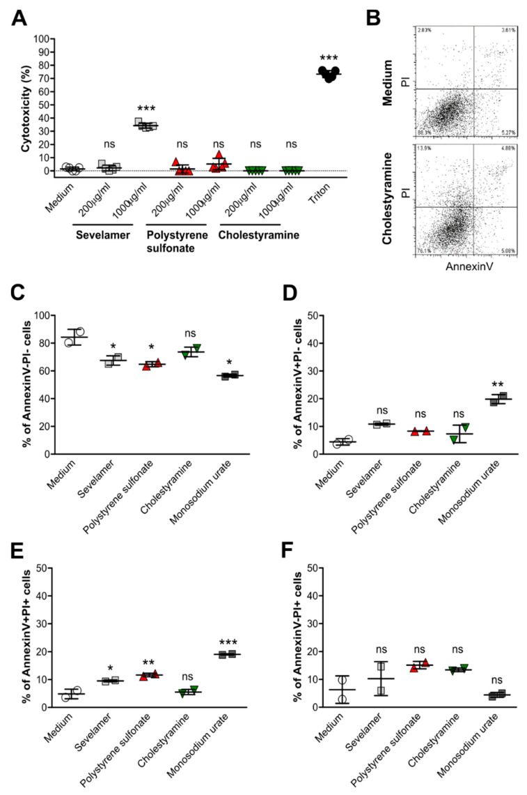
Figure 3.
Crystals of ion-exchange resins induce cell death in Caco2 intestinal epithelial cells. Human Caco2 intestinal epithelial cell lines were stimulated with different concentrations (200 or 1000 µg/mL) of sevelamer, polystyrene sulfonate, cholestyramine, or monosodium urate crystals for 24 h. Triton was used as a positive control. (A) After stimulation, culture supernatants were collected and lactate dehydrogenase (LDH) assays was performed. Cytotoxicity is presented as a percentage (%) (n = 5). (B–F) AnnexinV/propidium iodide (PI) staining of Caco2 cells was performed by flow cytometry (B). The percentage of live (AnnexinV-PI-) (C), apoptotic (AnnexinV+PI-) (D), late apoptotic/early necrotic (AnnexinV+PI+) (E), and necrotic (AnnexinV-PI+) (F) Caco2 cells after stimulation with 1000 µg/mL of different drug crystals was determined (n = 2). Data are means ± SD. * p ≤ 0.05; ** p ≤ 0.01; *** p ≤ 0.001; ns, not significant (p > 0.05) versus the medium using one-way ANOVA.

