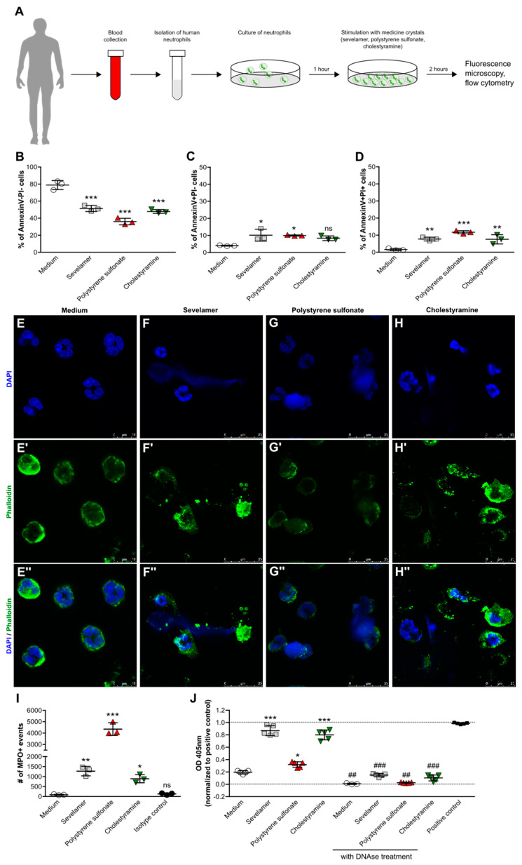
Figure 5.
Crystals of ion-exchange resins induce neutrophil extracellular trap formation. (A) Schematic showing work flow. Human blood neutrophils were isolated from healthy individuals and stimulated with or without sevelamer, polystyrene sulfonate, or cholestyramine crystals (1000 µg/mL) for 2 h. (B–D) After stimulation, the percentage of live (AnnexinV-PI-) (B), apoptotic (AnnexinV+PI-)(C), and late apoptotic/necrotic (AnnexinV+PI+) (D) neutrophils was quantified by flow cytometry (n = 3 from 3 donors). (E–H) Neutrophils were stained with DAPI (blue, stains nuclei) (E–H) and phalloidin (green, stains actin) (E’–H’) to visualize neutrophil extracellular trap formation in response to sevelamer (F), polystyrene sulfonate (G), and cholestyramine (H) crystals or the medium control (E). Images are also shown as a merge of DAPI and phalloidin (400× or 630× magnification (E’’–H’’)). (I) The number of myeloperoxidase (MPO)-positive events was determined in the supernatants of human neutrophils stimulated with or without sevelamer, polystyrene sulfonate, or cholestyramine crystals (1000 µg/mL) after 2 h by flow cytometry. (J) Quantification of released histone-complexed DNA fragments (OD 450 nm) in culture supernatants of crystal-stimulated neutrophils after 2 h using a cell death detection ELISAPLUS assay. DNAse treatment was used as the control. Data are means ± SD. * p ≤ 0.05; ** p ≤ 0.01; *** p ≤ 0.001; ns, not significant (p > 0.05) versus the medium, and ## p ≤ 0.01; ### p ≤ 0.001 versus without DNAse treatment, using one-way ANOVA.

