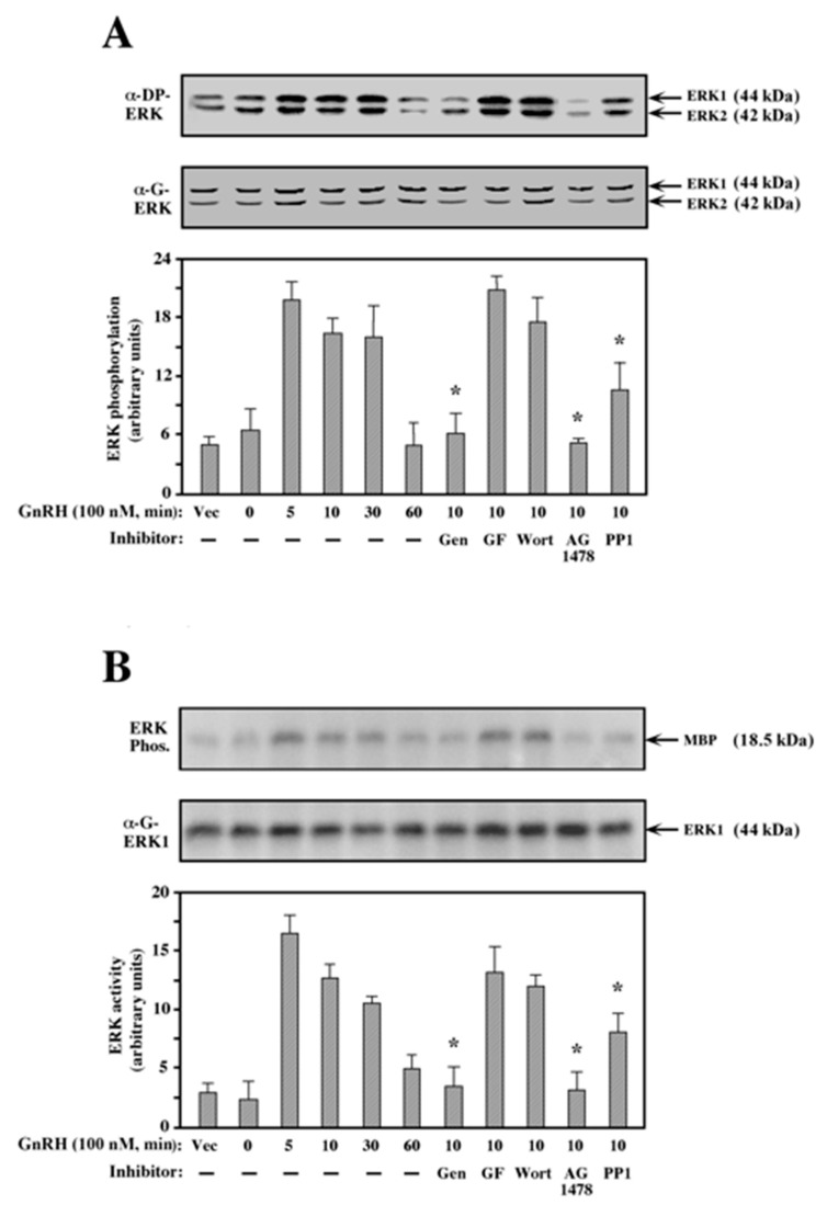
Figure 3.
Effect of various inhibitors on ERK activation by GnRH-a. (A) Phosphorylation of ERK: A plasmid-containing mouse GnRHR was transfected into COS7 cells using a DE-dextran technique (Materials and Methods), and the cells were treated as described in the legend to Figure 1. After serum starvation, the cells were either pretreated (15 min) with 200 μM genistein (Gen), 3 μM GF109203X (GF), 25 nM wortmannin (Wort), 5 μM AG1478, or 5 μM PP1, or left untreated. GnRH-a (10−7 M, 10 min) was added to the pretreated and untreated cells (5, 10, 30, and 60 min), or the cells were left untreated as a vector control (Vec. 0). Phosphorylation of ERK1 and ERK2 was detected with anti-DP-ERK antibody (α-DP-ERK). The total amounts of ERK1 and ERK2 were detected with the anti-ERK antibody (α-G-ERK). The results in the bar graphs below are an average of three experiments. Note: * p < 0.05. (B) Activation of ERK: The GnRHR-transfected COS7 cells were treated as in (A). The activity of ERK towards myelin basic protein (MBP) was determined (ERK Phos.) as described under the Methods section. The total amount of ERK was detected with the anti-ERK antibody (α-G-ERK); the site of ERK1 migration is indicated. The bar graphs below are an average of three experiments. Note: * p < 0.05.

