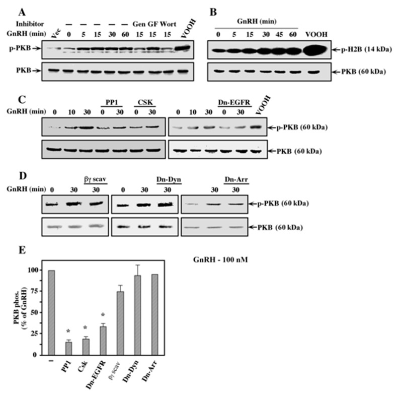
Figure 8.
Mechanism of activation of PI3K/PKB by GnRH-a. (A) COS7 cells were transfected with a plasmid-containing mouse GnRHR. Thirty-two hours after transfection, the cells were serum-starved for 16 h, after which the plates were pretreated with either 200 μM genistein (Gen), 3 μM GF109203X (GF), or 25 nM wortmannin (Wort). Then, the cells were either stimulated with GnRH-a (10−7 M for the indicated times) or with the non-specific activator peroxovanadate (200 µM H2O2, 100 µM vanadate; VOOH). Cellular extracts of these cells were subjected to a Western blot analysis using either anti-phospho-PKB antibody (Ser 473; p-PKB) or anti-PKB antibody (PKB). These results were reproduced twice. (B) COS7 cell were treated as in (A) and then subjected to immunoprecipitation with anti-PKB antibody followed by an in vitro phosphorylation reaction with histone H2B as a substrate (p-H2B). The phosphorylation was detected by autoradiography and the amount of immunoprecipitated PKB was detected with anti-PKB antibody (PKB). These results were reproduced twice. (C) COS7 cell were co-transfected with plasmid-containing mouse GnRHR together with plasmid-containing Csk, K721A-EGF receptor (Dn-EGFR), or no insertion as vector control. Thirty-two hours after transfection, the cells were serum-starved for 16 h, after which two plates were pretreated with the c-Src inhibitor PP1 (5 μM, 15 min). Then, the cells were stimulated with GnRH-a (10−7 M for the indicated times) and phosphorylation of PKB was detected with either anti-phospho PKB (p-PKB) or with anti-PKB (PKB) antibodies. These results were reproduced 3 times. (D) COS7 cells were co-transfected with mouse GnRHR, together with either CD8-tagged βARK (βγ scav), K44A-dynamin (Dn-Dyn), or V54D-β-arrestin2 (Dn-Arr), or no insertion as vector control. Two days after transfection, the cells were serum-starved for 16 h and then either treated with GnRH-a (10−7 M; 30 min) or left untreated (0). Activation of PKB was determined by Western blot analysis as in (C). The results were reproduced twice. (E) Activation of PKB was determined by densitometry. The bar graphs represent percentage activation from GnRH-a stimulated COS7 cells that were co-transfected with GnRHR and vector control in each experiment. The results represent averages and standard errors of 3 experiments. Note: * p < 0.05.

