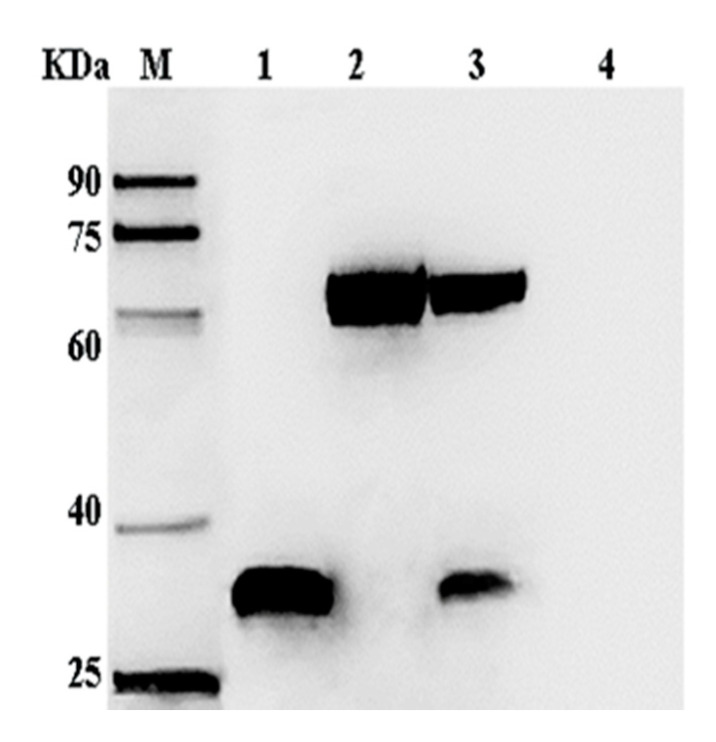
Figure 4.
Western blotting analysis of VP35 and VP4 expression in Sf-9 cells. Lane M, protein markers; lanes 1–3, Sf-9 at 72 h post-infection with recombinant viruses AcMNPV-VP35, AcMNPV-VP4, and AcMNPV-VP35-VP4, respectively; lane 4, Sf-9 at 72 h post-infection with wild virus. The primary antibody was mouse anti-His-tag, and the secondary antibody was HRP-conjugated goat anti-mouse IgG.

