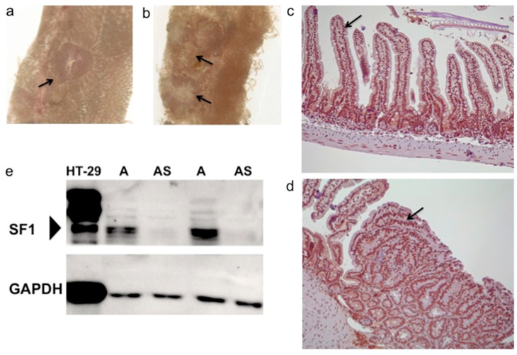
Figure 1.
SF1 expression in the intestine. (a,b) Representative polyps (arrow) in the small intestine of 4-month old ApcMin/+;Sf1+/− mice as viewed under the stereomicroscope. (c) Immunohistochemistry using anti-SF1 antibody of normal mouse intestine. Arrow indicates splicing factor 1 (SF1) expression in nuclei of intestinal villi and in (d) nuclei of adenoma cells of ApcMin/+ mice. (e) Immunoblotting using anti-SF1 antibody on cell lysate from HT-29 human colon cancer cells and lysates from ApcMin/+ (A) and ApcMin/+;Sf1+/− (AS) mouse intestines. (Bottom) GAPDH expression in the samples.

