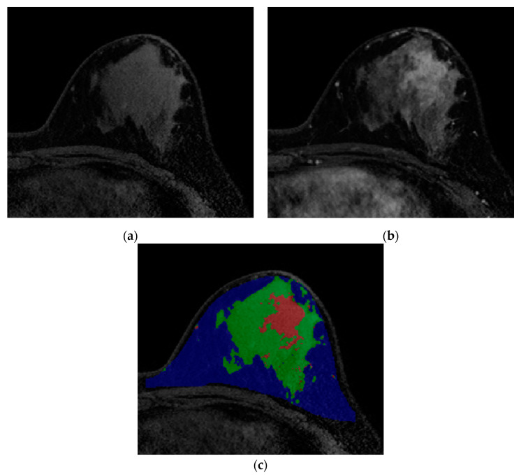
Figure 2.
Images from breast MRI of a 42-year-old woman with triple-negative invasive ductal carcinoma in the left breast. (a) Pre-enhanced T1-weighted axial image discriminates fat and non-fat regions. (b) Contrast-enhanced T1-weighted image shows a 4 cm malignant mass. At this stage, differentiation between tumor and normal fibroglandular tissue is conducted. (c) A color-overlay image showing differentiation between fat (blue), normal fibroglandular tissue (green), and tumor (red). In this patient, the tumor-fat interface volume was 1.80 cm3 and classified as low interface group. Final pathology after surgery revealed a pCR.

