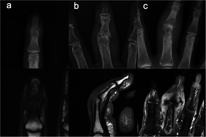
Fig. 1.
a The right index finger of a 35-year-old man. The distal interphalangeal joint shows joint space narrowing in a simple radiograph, while T1 weighted image shows intramedullary marked low signal intensity, indicating osteomyelitis. b The right long finger of an 80-year-old woman. The proximal interphalangeal joint shows joint space narrowing and radial angulation deformity in a simple radiograph; however, a normal marrow signal is shown in the T1 weighted image. c The right long finger of a 58-year-old woman. The proximal interphalangeal joint shows definite osteolysis in a simple radiograph. The proximal phalanx reveals extensive intraosseous abscess in contrast-enhanced T1 weighted image

