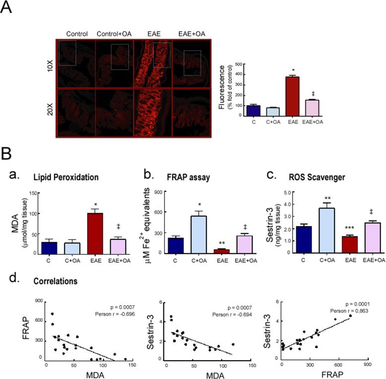
Fig. 2.
OA treatment reduces oxidative stress in colon tissue from EAE mice. Representative photomicrographs of a colon tissue stained with DHE. Histological analysis by fluorescence microscopy and quantification. Objective lens × 10 and × 20. b Expression levels in colon of (a) malondialdehyde, MDA, (b) ferric reducing/antioxidant power, FRAP, and (c) the ROS disruptor Sestrin-3. (c, d) Scatter plots of the correlation between oxidative parameters. Results were expressed as the mean ± SEM, n = 5–7 per group. *p < 0.001, **p < 0.01, and ***p < 0.05 vs control; and ‡p < 0.001 vs untreated-EAE. C, healthy mice. C + OA, healthy mice treated with OA. EAE, induced mice. EAE + OA, induced-mice treated with OA

